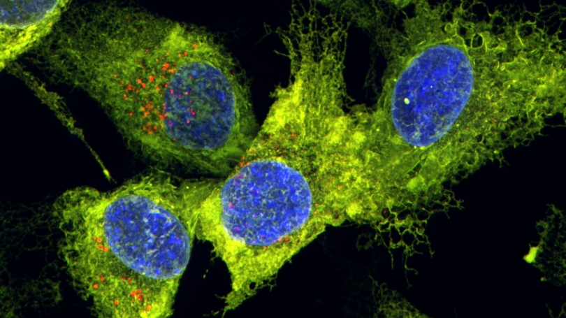
Dual labeled (red and green) endoplasmic reticulum (ER) in a human breast cancer cell line. Nuclei are stained blue. Red puncta mark sites of lysosomal engulfment of the ER. Image acquired by Sevdora Eshnazarova (Dartmouth '25, Shoemaker Lab) on the Nikon SoRa Spinning Disk Confocal.
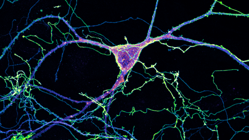
Primary hippocampal neuron expressing a glutamate-sensitive fluorescent reporter (iGluSnFR3, blue/green) and an ER lumen marker (mcherry-ER, magenta). Image acquired by Cam Paton (MCB, Hoppa Lab) on the Nikon SoRa Spinning Disk Confocal.
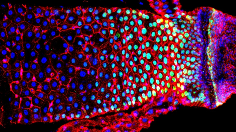
Drosophila adult intestine, midgut-hindgut boundary (right). Cell junctions (red), nuclei (blue), and cells in which the Wnt signal transduction pathway is active (green). Image acquired by Hassina Benchabane (Research Scientist, Ahmed Lab) on the Nikon A1R Laser Scanning Confocal.
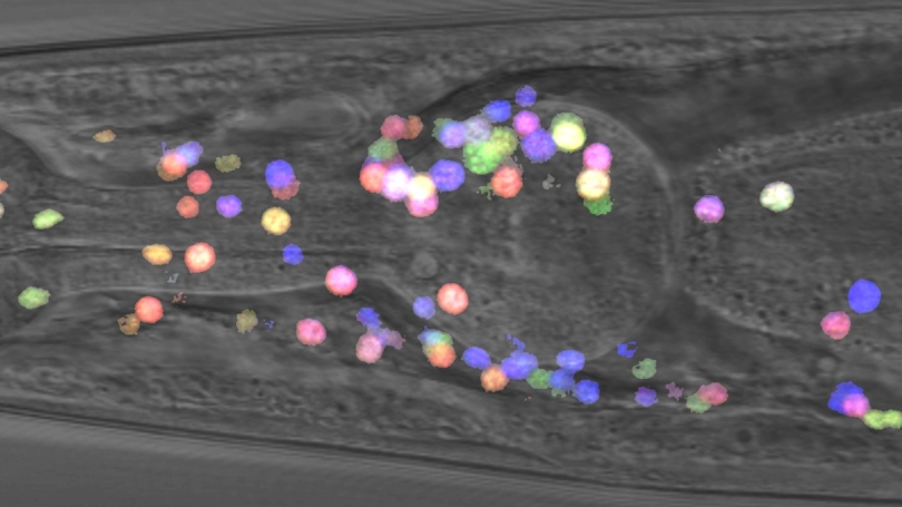
C. elegans head imaged with differential interference contrast (DIC). The NeuroPAL multicolor fluorescent reporter system is used to determine neuronal identity based on color and position. Image acquired by Kelli Cannon (Postdoctoral Researcher, Ghosh Lab) on the Zeiss LSM 880 Laser Scanning Confocal.
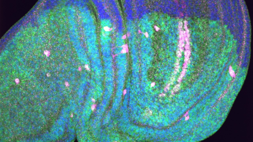
E3 ubiquitin ligase (Ctrip, green), Wingless target gene reporter (Scarlet-Senseless, magenta), and nuclei (blue) in the Drosophila larval wing disc. Image acquired by Kai Yuan (MCB, Ahmed Lab) on the Nikon Spinning Disk Confocal.
- Undergraduate
- Graduate
- Research
- Foreign Study Program
- Seminars, News & Events
- People
- SEPA
Back to Top Nav
Back to Top Nav
Back to Top Nav
Back to Top Nav